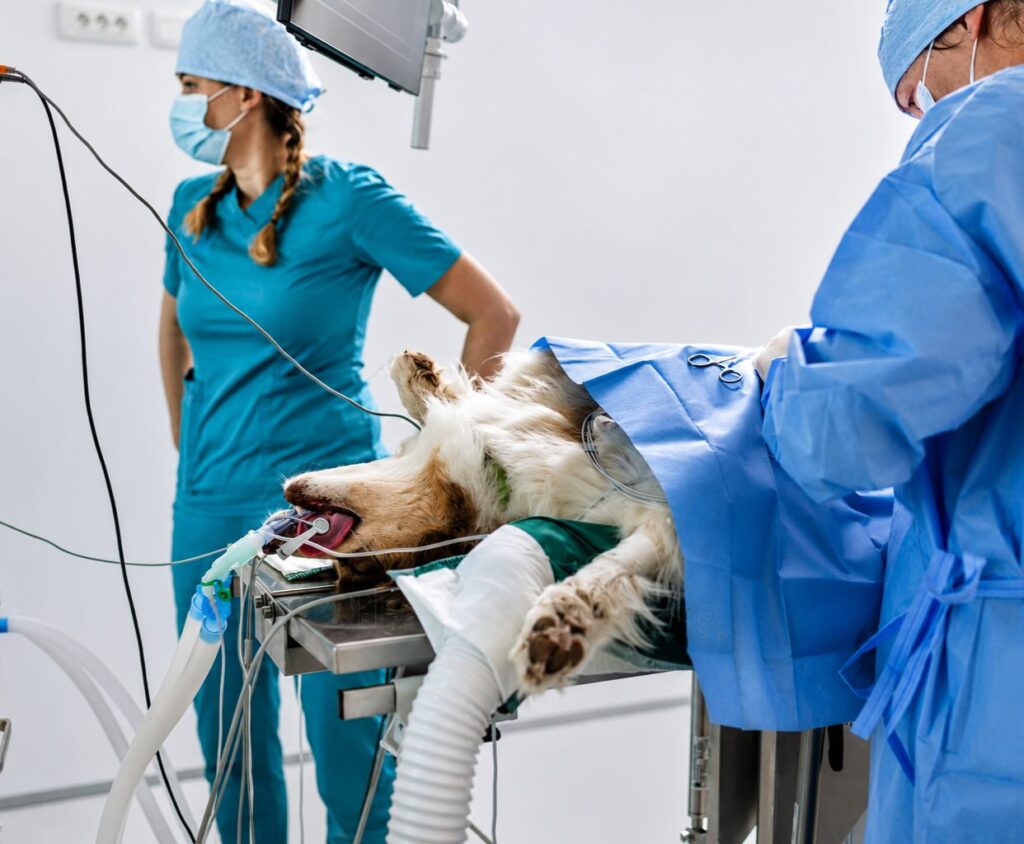
A cancer diagnosis in a pet raises an enormous number of questions all at once. What kind of tumor is it? Has it spread? What are the options? And if surgery is recommended, what does that actually involve? For many owners, the idea of their companion undergoing an operation is deeply unsettling, and the terminology of pet oncology surgery can make it feel even more overwhelming. Understanding what tumor removal surgery for pets entails, what the decision-making process looks like and what recovery involves gives owners the clarity they need to make informed choices with their veterinary team.
Cancer is one of the most frequently encountered serious conditions in older pets, and surgery remains one of the most effective treatments available across many tumor types. Pet cancer does not always develop with obvious warning signs in the early stages, which is why knowing signs of cancer emergencies in pets helps owners recognize when a lump, a behavioral change or a sudden decline in condition needs immediate professional attention rather than delayed investigation.
This guide covers why surgery is used, how the decision is made, what happens in the operating room, what recovery looks like and when pet cancer becomes an acute emergency.
Why Surgery Is Used to Treat Pet Cancer
Surgery remains one of the most effective tools in pet cancer treatment. For many tumor types, complete surgical removal offers the best chance of a cure or a meaningful extension of quality life. Unlike systemic therapies that affect the entire body, surgery removes the tumor directly and, when performed with adequate margins, eliminates local disease entirely.
Surgery is often used alongside other therapies rather than in isolation. Some tumors are treated with surgery followed by additional targeted treatment to address microscopic residual disease. Others are debulked surgically to enhance the effectiveness of radiation. Your veterinary team will discuss which combination of approaches gives the best result for your pet’s specific tumor type.
Goals of Pet Tumor Removal
Not all pet tumor removal surgeries have the same objective. Understanding which goal applies to your pet’s situation shapes everything from how aggressive the surgery is to what the expected outcome looks like.
Curative intent surgery aims to remove all cancerous tissue with clean margins, meaning the entire tumor is excised along with a surrounding border of normal tissue. When clean margins are achieved and the tumor has not spread to other areas of the body, the pet may be effectively cured of that cancer. Debulking surgery aims to remove as much of the tumor as possible without the expectation that all cancer cells can be eliminated. This approach is used when complete removal is not technically feasible, or when reducing the tumor burden will make other treatments more effective. Palliative surgery does not address the tumor itself but relieves the symptoms it is causing, with the goal being comfort and improved day-to-day function rather than tumor control.
Making the Decision for Pet Tumor Removal
The decision to proceed with tumor removal surgery for pets involves multiple layers of information gathered through a diagnostic workup. It is never made based on finding a lump alone.
Biopsy and Tumor Typing
Before any surgical plan is finalized, knowing what type of tumor is present is essential. A fine needle aspirate may provide initial information, but a full biopsy with histopathology is required in most cases to confirm the diagnosis and characterize the tumor’s behavior. The grade of the tumor, meaning how abnormal the cells appear and how aggressively they are likely to behave, directly influences the surgical approach. A low-grade tumor may be managed with a conservative excision while a high-grade tumor requires much wider margins and may warrant additional therapies after surgery. Our guides on what causes mast cell tumors in cats and what does a skin tumor look like on a dog explain how specific surface tumors are evaluated before surgery is discussed.
Staging
Staging evaluates how far the cancer has spread beyond the primary tumor. This is a critical step in pet oncology surgery planning because operating on a localized tumor when cancer has already spread to distant organs may not change the prognosis significantly and may expose the pet to surgical risk without meaningful benefit.
Staging typically involves blood work, urinalysis, regional lymph node evaluation, chest X-rays to look for lung metastasis and abdominal ultrasound to evaluate the liver, spleen and other organs. Advanced imaging such as CT scanning provides more detailed information about tumor boundaries and lymph node involvement, particularly for tumors in complex anatomical locations.
Overall Health Assessment
The pet’s overall health determines their ability to safely undergo anesthesia and surgery and their capacity to recover. Blood work assessing organ function, a cardiovascular evaluation and a review of any current health conditions are all part of pre-surgical assessment. Older pets or those with concurrent illnesses can still undergo pet tumor removal, but these factors require careful anesthetic planning and sometimes additional pre-operative preparation.
Types of Pet Cancer That Commonly Require Surgery
Skin and Subcutaneous Tumors
Skin tumors are among the most frequently surgically treated in both dogs and cats. Mast cell tumors, soft tissue sarcomas, squamous cell carcinomas and various benign tumors all commonly require excision. The surgical approach and margin requirements vary considerably by tumor type. Mast cell tumors require wide lateral and deep margins because of their infiltrative nature, while a benign lipoma may be removed with minimal margins. Because the post-operative wound site in these cases needs careful monitoring, owners should understand the basics of wound care. The guide on how to properly clean a cat wound and prevent infection provides practical guidance applicable to any post-surgical wound care at home.
Splenic Tumors
Splenic masses are common in older dogs and represent one of the more urgent surgical scenarios in veterinary medicine. The spleen can harbor both benign and malignant masses, and because it has a rich blood supply, splenic tumors are prone to sudden rupture causing rapid internal hemorrhage. Splenectomy is both diagnostic and therapeutic. The removed spleen is submitted for histopathology to determine whether the mass was benign or malignant, which guides decisions about follow-up monitoring or additional treatment.
Mammary Tumors
Mammary gland tumors are one of the most common forms of pet cancer in intact female dogs and are also seen in female cats. Surgical removal is the primary treatment and may involve removing the affected gland alone or a chain of glands depending on the extent of disease. The prognosis after surgery depends on the histopathological findings including tumor type, grade and whether lymph node involvement is present.
Intestinal and Abdominal Masses
Tumors of the gastrointestinal tract, liver and other abdominal organs may require surgery when they are causing obstruction, hemorrhage or significant symptoms. Intestinal lymphoma in cats may be diagnosed through full-thickness biopsy obtained during exploratory surgery, and certain localized intestinal tumors can be treated with resection and anastomosis. For a broader understanding of how lymphoma develops in cats and what treatment involves beyond surgery, the guide on what causes lymphoma in cats provides important background context.
Bone Tumors
Bone tumors, particularly osteosarcoma in large breed dogs, require the most extensive surgical intervention. Limb-sparing procedures or amputation may be discussed depending on the location and extent of the tumor. These surgeries are typically performed at specialist referral centers by veterinary surgical oncologists and are often combined with systemic therapy.
Nasal and Oral Tumors
Tumors in the nasal cavity and oral cavity can involve complex anatomy and may affect the ability to eat, breathe and smell. Surgery for these tumors often requires significant tissue removal and reconstructive planning and may be combined with radiation therapy for optimal results.
Comparing Surgical Goals in Pet Oncology Surgery
| Surgical Goal | When It Applies | Expected Outcome | Additional Therapy Often Needed |
| Curative intent | Localized tumor, no evidence of spread, complete excision possible | Possible cure if clean margins achieved | Sometimes, depending on tumor type |
| Debulking | Tumor too large or complex for full removal | Reduced tumor burden, improved quality of life | Yes, usually combined with other treatments |
| Palliative | Symptom relief when cure is not the goal | Improved comfort and function | Supportive care focused approach |
What Happens During Tumor Removal Surgery
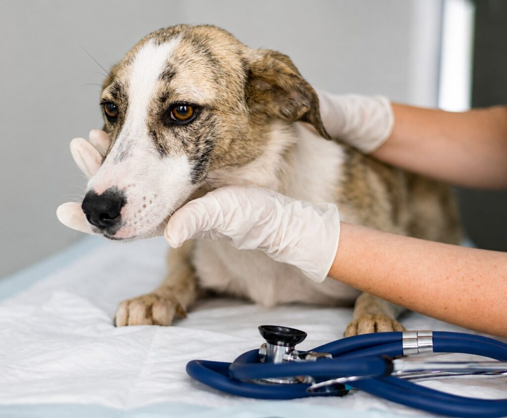
Anesthesia and Preparation
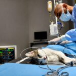
The pet is fasted before anesthesia and receives an intravenous catheter for fluid support and medication delivery. A pre-anesthetic examination confirms the pet is stable for the procedure. Monitoring equipment including electrocardiogram, blood pressure, pulse oximetry and capnography tracks vital signs continuously throughout the surgery.
Surgical Margins
The concept of surgical margins is central to successful pet tumor removal. When a surgeon removes a tumor, they do not cut along its visible edge. They remove the tumor plus a surrounding border of normal-appearing tissue, the width of which is determined by the tumor type and grade. After surgery, the removed tissue is submitted to a pathologist who examines the edges of the specimen to determine whether cancer cells reach the cut edge, called a positive or dirty margin, or whether cancer-free tissue surrounds the entire specimen, called a clean or negative margin. Clean margins are the goal and are the strongest indicator of successful local tumor control.
Lymph Node Assessment
Depending on the tumor type, regional lymph nodes may be sampled during surgery to determine whether cancer has spread beyond the primary site. This information influences post-surgical treatment recommendations and provides important prognostic information for the owner and veterinary team going forward.
Duration and Complexity
The length of tumor removal surgery varies enormously. A straightforward skin tumor removal may take under an hour. A splenectomy, intestinal resection or complex nasal tumor surgery can take several hours and involves more intensive monitoring and recovery support. The surgeon will discuss expected procedure length and any specific intraoperative risks with the owner before proceeding.
Post-Surgical Recovery
Immediate Post-Operative Period
After surgery, the pet is monitored closely during recovery from anesthesia. Pain management is a priority and is achieved through a combination of medications determined by the veterinarian rather than any single drug. Intravenous fluids continue until the pet is stable, eating and drinking. Most pets stay in hospital for at least one night after tumor removal surgery to allow for close monitoring of vital signs and wound integrity.
Wound Care and Activity Restriction
At home, the surgical incision needs to be kept clean, dry and observed daily for signs of infection, swelling or breakdown. An Elizabethan collar prevents the pet from licking or chewing at the incision site, which is one of the most common causes of wound complications. Activity restriction, meaning no running, jumping or rough play, is required for a minimum of two weeks for most soft tissue surgeries and longer for bone or reconstructive procedures.
Because dogs that have experienced significant blood loss before or during surgery may also face anemia in recovery, it is worth understanding what are symptoms of anemia in dogs so owners can recognize if pale gums, weakness or unusual fatigue during the recovery period warrants a call back to the veterinary team.
Histopathology Results
The pathology report from the removed tumor arrives typically within five to ten days after surgery. This report provides the definitive diagnosis, grade and margin assessment that guide all subsequent decisions. If margins are incomplete, a second surgery to remove additional tissue may be recommended. If the tumor type carries a significant risk of spread, additional staging or therapies are discussed at the follow-up appointment.
Follow-Up Monitoring
After recovery from pet oncology surgery, a monitoring schedule is established based on tumor type and grade. Follow-up visits involve physical examination, lymph node assessment and imaging at intervals determined by recurrence risk. Early detection of recurrence gives the most options for additional treatment and is a key reason why owners should maintain consistent recheck appointments even when the pet appears to be doing well.
When Tumor Surgery Becomes an Emergency
Most tumor removal surgeries are planned procedures following diagnosis and staging. However, certain situations convert pet cancer into an emergency requiring immediate intervention without the opportunity for advance planning.
A ruptured splenic tumor causing internal hemorrhage is one of the most common emergency tumor surgeries in dogs. A pet that collapses suddenly with pale gums and a distended abdomen may have a splenic mass that has bled into the abdominal cavity and requires emergency splenectomy. Dogs with splenic rupture can deteriorate within one to two hours, and the dog internal bleeding symptoms resource outlines exactly what to watch for and how quickly to act when these signs appear.
In these situations, understanding the impact of delayed emergency care is critical because the window for successful stabilization and surgery narrows rapidly as blood loss progresses and the pet enters shock.
Questions Worth Asking Your Veterinary Team
Before agreeing to tumor removal surgery for pets, owners benefit from having clear answers to several key questions. What type of tumor is this and how does it typically behave? Is this surgery intended to be curative or will additional treatment be needed? What are the expected surgical margins and how will we know if they were achieved? What is the recovery timeline and what should I watch for at home? Are there non-surgical options and what would happen if we chose them? What does the follow-up monitoring schedule look like after discharge?
Good communication with the veterinary team ensures that the surgical decision aligns with the owner’s goals for their pet’s quality of life and that expectations for outcome are realistic and clearly understood by everyone involved.
Giving Your Pet the Best Chance
Tumor removal surgery for pets is a significant undertaking, but for many animals it represents the most effective path to controlling or eliminating pet cancer and restoring good quality of life. The process from diagnosis through surgery and recovery is a partnership between owner and veterinary team. Informed owners who understand each step are better equipped to make good decisions, recognize early warning signs during recovery and provide the best possible support to their pet throughout the entire process.
At North MS Pet Emergency, we provide after-hours and weekend emergency care for pets experiencing acute cancer-related complications including internal hemorrhage, respiratory distress and collapse from tumor-related causes. If your pet is showing signs of an urgent pet cancer emergency, do not wait for a scheduled appointment. Contact us immediately for emergency evaluation and stabilization.
FAQs
Q: What does tumor removal surgery for pets actually involve?
A: The surgeon removes the tumor plus a border of surrounding healthy tissue called a margin. The removed specimen is sent for pathology to confirm the diagnosis and assess margin cleanliness. Recovery involves wound care, activity restriction and a follow-up pathology report guiding next steps.
Q: How do vets decide if pet tumor removal surgery is the right option?
A: The decision depends on tumor type, grade, location and staging results. Blood work, imaging and biopsy are completed before surgery is recommended. The pet’s overall health and the owner’s quality-of-life goals also significantly influence the treatment recommendation that is made.
Q: What are clean margins and why do they matter in pet oncology surgery?
A: Clean margins mean cancer-free tissue surrounds the entire tumor specimen after removal, indicating the local tumor was fully excised. Dirty margins mean cancer cells reach the cut edge, increasing recurrence risk and potentially prompting a second surgery or additional targeted therapy.
Q: How long does recovery from pet cancer surgery take?
A: Most soft tissue tumor removals require two weeks of activity restriction and wound care at home. Pathology results return within ten days. More complex surgeries involving bone, abdominal organs or reconstruction require longer recovery periods and more intensive post-operative monitoring.
Q: When does a pet cancer situation become an emergency?
A: Sudden collapse, pale gums, rapid breathing or a distended abdomen in a pet with a known or suspected tumor can indicate a ruptured mass causing internal bleeding. This is a surgical emergency requiring immediate veterinary care rather than waiting for any planned appointment.
Recent Posts
-
 Why Cats Suddenly Lose Use of Their Back Legs From a Blood Clot
 -
 Why Are Lilies Poisonous to Cats and How Fast Symptoms Can Appear
 -
 What Are the Signs of Pyometra in Dogs and Why It Cannot Wait
 -
 How To Recognize Warning Signs In A Pet In Pregnancy
 -
 Why Some Pet Emergencies Require Overnight Pet Care And Monitoring