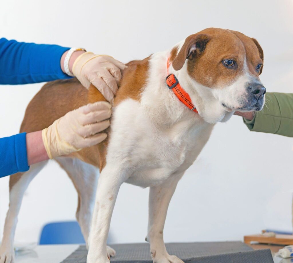
Finding a lump on your dog during a routine pat or grooming session is one of those moments that can send immediate worry through any owner. The question that follows almost instinctively is whether it is serious. The honest answer is that skin growths on dogs range from completely harmless to medically significant, and appearance alone is not enough to tell them apart. Understanding what does a skin tumor look like on a dog, which characteristics are more concerning than others and when the situation calls for a pets emergency evaluation helps you respond appropriately rather than either dismissing something that needs attention or panicking unnecessarily.
This guide covers the most common types of dog skin tumors, how to describe what you are seeing, what veterinary diagnosis involves and when urgency is warranted.
Why Dogs Develop Skin Growths
The skin is the largest organ in the body and contains a wide variety of cell types, each of which can proliferate abnormally to form a tumor. Dogs are actually more prone to developing skin and subcutaneous tumors than most other domestic animals, and skin tumors make up a significant proportion of all canine cancer diagnoses.
Not all skin growths are cancerous. The word tumor simply refers to an abnormal mass of cells and does not inherently indicate malignancy. Many dog skin tumors are benign, meaning they do not invade surrounding tissue or spread to other areas of the body. Others are malignant and require prompt treatment to prevent progression. The challenge is that visual inspection alone cannot reliably distinguish between them.
Risk Factors for Dog Skin Tumors
Age is the most consistent risk factor. Most skin tumors are diagnosed in middle-aged to older dogs, though certain types can appear in younger animals. Sun exposure contributes to certain tumor types, particularly in dogs with light-colored or thin coats and areas of minimal pigmentation such as the abdomen and nose. Breed predisposition is significant for several tumor types, and chronic skin irritation or inflammation at a specific site has been associated with tumor development over time in some cases.
What Does a Skin Tumor Look Like on a Dog?
The appearance of skin growths on dogs varies enormously depending on the cell type of origin, the location and whether the growth is benign or malignant. There is no single appearance that all tumors share, which is one reason why veterinary evaluation is the only reliable way to determine what a growth is.
General Characteristics to Observe
When you find a lump on your dog, these are the features worth noting before your veterinary appointment. Size and shape matter, as does whether the edges of the growth are well-defined or irregular. Note whether the surface is smooth, rough, ulcerated or crusted. Observe whether the lump appears to sit just beneath the skin surface and moves freely when pressed, or whether it feels firmly attached to underlying tissue. Color is worth noting too, including whether the growth is the same color as surrounding skin, darker, red or multicolored.
Equally important is noting how long the lump has been there, whether it has changed in size, appearance or texture over time and whether the dog appears to notice it, lick it or scratch at it.
Common Types of Dog Skin Tumors
Lipomas
Lipomas are benign tumors composed of fat cells and are among the most frequently encountered skin growths on dogs. They typically feel soft, smooth and slightly squishy to the touch and move freely under the skin when pressed. They are most common in middle-aged and older dogs, particularly in overweight individuals, and tend to appear on the trunk, upper legs and armpits. Lipomas are usually slow-growing and non-painful.
While lipomas are generally harmless, not every soft fatty-feeling lump is a lipoma. A variant called an infiltrative lipoma grows into surrounding muscle tissue and is more problematic. Fine needle aspirate is needed to confirm the diagnosis and distinguish a lipoma from other soft tissue masses.
Mast Cell Tumors
Mast cell tumors are one of the most common and clinically significant types of dog skin tumors. They are notoriously variable in appearance, which is why they are sometimes referred to as the great imitators in veterinary dermatology. A mast cell tumor might look like a soft raised lump, a firm nodule, an inflamed red area, an ulcerated lesion or something that closely resembles an insect bite. It may change in size from day to day due to the release of inflammatory chemicals from the tumor cells.
Mast cell tumors are graded based on how aggressive they appear under the microscope, and this grade significantly influences treatment decisions and prognosis. They are most commonly diagnosed in Boxers, Boston Terriers, Bulldogs, Labrador Retrievers, Golden Retrievers and Pugs, among other breeds. Any suspicious skin growth in these breeds warrants prompt veterinary evaluation.
Sebaceous Cysts and Adenomas
Sebaceous glands in the skin produce oil that lubricates the coat. Blockages in these glands can produce cysts, which are fluid-filled sacs beneath the skin surface that feel firm or fluctuant depending on their contents. Sebaceous adenomas are benign overgrowths of these glands that often appear as small warty or cauliflower-like growths, most commonly on the head, eyelids and back, particularly in older dogs and certain breeds including Cocker Spaniels and Poodles.
These are generally benign, though they can become irritated, rupture or develop secondary infections. A ruptured cyst may look inflamed and discharge a thick material. Read more about recognizing and treating pet skin infections if you notice signs of infection developing around any skin growth.
Histiocytomas
Histiocytomas are benign tumors of immune cells that appear most frequently in young dogs under three years of age. They typically present as small, raised, button-like hairless red growths that appear suddenly and grow quickly over a few weeks. Despite their rapid onset and distinctive red appearance, most histiocytomas regress spontaneously within one to three months without treatment. Veterinary confirmation is still recommended to rule out more serious diagnoses that can look similar.
Squamous Cell Carcinoma
Squamous cell carcinoma is a malignant tumor of the skin’s surface cells. It often develops on areas with less pigmentation or areas exposed to chronic sun damage, including the nose, lips, abdomen and ear tips. Squamous cell carcinoma typically appears as a firm, raised, often ulcerated or crusty lesion that does not heal. It may bleed intermittently. This tumor type can invade surrounding tissue and in some cases spread to regional lymph nodes. Early diagnosis and surgical removal while the lesion is still small significantly improves outcomes.
Melanoma
Melanomas in dogs can affect both pigmented and non-pigmented skin. Benign melanocytomas are common and may appear as dark, well-circumscribed raised growths on haired skin. Malignant melanoma most often arises in the mouth, nail beds and mucocutaneous junctions but can occasionally appear on haired skin as well. A dark or black growth that is growing, irregular in shape or located near the mouth, lips or a nail bed warrants prompt veterinary evaluation.
Soft Tissue Sarcomas
Soft tissue sarcomas are a group of malignant tumors arising from connective tissue beneath the skin. They often appear as firm, deep-seated masses that feel attached to underlying tissue rather than freely movable. They may grow slowly at first and are not always painful in the early stages. However, they can invade local tissue significantly and some types have the potential to spread to internal organs. Any firm deep lump that does not move freely should be evaluated without delay.
Comparing Common Dog Skin Tumor Types
| Tumor Type | Typical Appearance | Behavior | Urgency of Evaluation |
| Lipoma | Soft, moveable, smooth | Benign, slow-growing | Routine but confirm with aspirate |
| Mast cell tumor | Variable, may change size daily | Potentially aggressive | Prompt evaluation needed |
| Histiocytoma | Small raised red button, hairless | Benign, usually self-resolves | Confirm diagnosis to rule out other causes |
| Sebaceous cyst or adenoma | Firm or fluctuant, warty surface | Benign, may get infected | Routine unless infected or growing rapidly |
| Squamous cell carcinoma | Ulcerated, crusty, non-healing lesion | Malignant, locally invasive | Urgent evaluation recommended |
| Soft tissue sarcoma | Firm, deep, attached to underlying tissue | Malignant, locally invasive | Prompt evaluation needed |
| Melanoma | Dark, raised, irregular in some cases | Variable, location-dependent | Prompt evaluation if near mouth or nail bed |
When Should You Be More Concerned?
Certain features of a skin growth increase the level of concern and should prompt a sooner rather than later veterinary appointment. A lump that has changed size, shape or texture over a short period is worth evaluating promptly. Any growth that is ulcerated, bleeding, crusting or oozing needs to be seen. A mass that appears to be growing into rather than resting on top of the surrounding tissue warrants investigation. Multiple new lumps appearing simultaneously, or a growth in a location such as near the mouth, a nail bed or on the eyelid, should be assessed without long delay.
In a dog with a previously diagnosed cancer, any new growth or change to an existing mass should be reported to the veterinary team. Understanding signs of cancer emergencies in pets provides broader context on when a cancer-related situation crosses into urgent territory.
How Are Dog Skin Tumors Diagnosed?
Fine Needle Aspirate
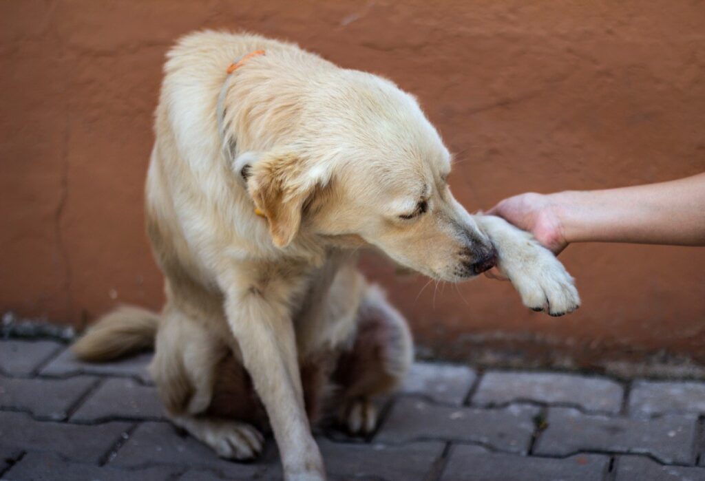
A fine needle aspirate is the first and most accessible diagnostic step for most skin growths. A small needle is inserted into the mass to collect cells, which are then examined under a microscope. This procedure is quick and usually does not require sedation. It provides a useful initial picture of what types of cells are present, though it cannot always give a complete answer on its own.
Biopsy and Histopathology
A tissue biopsy, where a larger sample is removed and processed for histopathology, provides the definitive diagnosis. It allows the pathologist to assess not only the cell type but also the degree of malignancy, the adequacy of surgical margins if a mass has been removed and features that predict biological behavior. This is the gold standard for characterizing any tumor type.
Staging
For tumors that are confirmed as malignant, staging tests determine whether the disease has spread beyond the primary site. These may include lymph node evaluation, chest X-rays and abdominal ultrasound. Staging guides treatment planning and gives the most accurate prognostic information.
Treatment Options for Dog Skin Tumors
Treatment depends entirely on what the growth is. Benign tumors that are not causing discomfort, are not in a problematic location and are not growing may be monitored over time with periodic recheck measurements. Surgical removal is the primary treatment for most malignant skin tumors and for benign growths that are causing irritation, infection or interfering with normal function.
For malignant tumors, additional therapies may be recommended alongside or following surgery depending on the tumor type and grade. Your veterinarian will guide treatment decisions based on the histopathology results and the dog’s overall health. Palliative options focused on comfort and quality of life are always discussed with owners when curative treatment is not the goal.
Post-surgical monitoring of the incision site is important. If you notice increasing swelling, discharge or failure of the wound to close normally, contact your veterinarian promptly rather than assuming it is part of normal healing.
Is This a Pets Emergency?
Most skin growths do not present as acute pets in emergency situations, but several scenarios require immediate veterinary attention rather than a routine appointment.
Seek emergency care if a tumor has ruptured and is actively bleeding and cannot be controlled with gentle pressure. If a mass near the throat or chest is causing visible difficulty breathing or swallowing, this is urgent. If a dog with a known mast cell tumor develops sudden systemic signs including facial swelling, vomiting, collapse or hives following handling of the tumor, this suggests a mass degranulation event requiring immediate evaluation. Any new growth that appears alongside signs of general illness such as lethargy, pale gums, loss of appetite or collapse is a combination that should not wait.
Understanding the impact of delayed emergency care makes clear why acting promptly in these moments rather than waiting for a scheduled slot can make a meaningful clinical difference.
What Every Dog Owner Should Take Away
Skin growths in dogs are common, and many are benign. But the only way to know for certain what a growth is and how it is likely to behave is through veterinary diagnosis. The question of what does a skin tumor look like on a dog does not have a single clean answer, because dog skin tumors present in many different ways and appearance alone is not a reliable guide to seriousness. Finding a new lump on your dog is a prompt to make a veterinary appointment, not a reason for immediate panic, but also not something to watch indefinitely without investigation.
At North MS Pet Emergency, we are available after hours and throughout weekends to evaluate dogs presenting with concerning skin growths or related acute complications. If your dog is showing signs of a pets emergency alongside a skin mass, do not delay seeking care. Contact us immediately for emergency evaluation and support.
FAQs
Q: What does a skin tumor look like on a dog?
A: There is no single appearance. Dog skin tumors can be soft or firm, smooth or rough, raised or flat, pigmented or skin-colored. Some ulcerate while others remain intact. Appearance alone cannot confirm whether a growth is benign or malignant, which is why veterinary diagnosis is always needed.
Q: Are all skin growths on dogs cancerous?
A: No. Many skin growths on dogs are benign, including lipomas, sebaceous cysts and histiocytomas. However, some malignant tumor types can look harmless at first glance. The only reliable way to distinguish benign from malignant is through a fine needle aspirate or biopsy performed by a veterinarian.
Q: How quickly should I have a new lump on my dog evaluated?
A: Any new growth that is ulcerated, bleeding, growing rapidly, changing appearance or located near the mouth, eye or nail bed should be seen within days. A soft stable lump that has been present without change for weeks can be evaluated at the next available routine appointment.
Q: What breeds are most at risk for dog skin tumors?
A: Boxers, Bulldogs, Golden Retrievers, Labrador Retrievers, Cocker Spaniels, Poodles and Schnauzers are among the breeds more frequently diagnosed with various types of skin tumors. However, any breed can develop skin growths, particularly as they age into their senior years.
Q: When is a skin tumor on a dog a pets emergency?
A: Seek immediate care if the tumor is actively bleeding without stopping, is causing breathing or swallowing difficulty, or if the dog develops systemic signs alongside a known mass such as collapse, pale gums or facial swelling. These situations require emergency evaluation without delay.
Recent Posts
-
 Why Are Lilies Poisonous to Cats and How Fast Symptoms Can Appear
 -
 What Are the Signs of Pyometra in Dogs and Why It Cannot Wait
 -
 How To Recognize Warning Signs In A Pet In Pregnancy
 -
 Why Some Pet Emergencies Require Overnight Pet Care And Monitoring
 -
 Understanding Tumor Removal Surgery for Pets and What It Involves